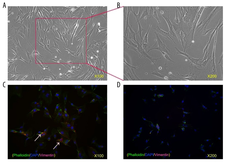
Figure 1.
Cultivation and identification of hPDLCs. (A) Fifth passage of hPDLCs. Cells were arranged in a sarciniform or swirl pattern and were fusiform in shape. Original magnification, 100×. (B) Fifth passage of hPDLCs. Original magnification, 200×. (C) Immunofluorescence strains of hPDLCs. Vimentin was found in the cytoplasm with a red color whereas keratin was not found in hPDLCs, indicating hPDLCs are mesenchymal cells derived from the embryonic mesoderm. Original magnification, 100×. (D) Immunofluorescence strains of hPDLCs. Original magnification, 200×.

