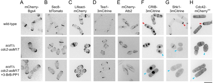
Fig. 1.
Polarized growth of scd1Δ cells during extended interphase. (A-H) Cell morphology and localization of polarity-associated proteins in cells of the indicated genotypes. 3-BrB-PP1 was added 5 h before imaging to inhibit analog-sensitive Cdc2 (bottom row). Arrowheads in F and G indicate detection (red) or no significant detection (blue) at cell tips. Arrowheads in H indicate enrichment (red) or no enrichment (blue) at tips. Scale bar: 10 µm. See also Fig. S1.

