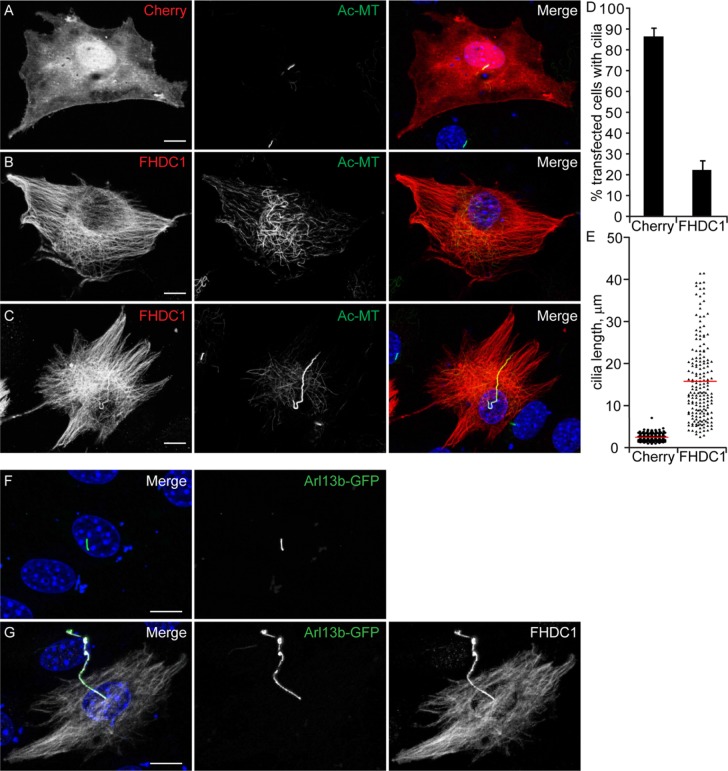
FIGURE 1:
FHDC1 expression induces cilia elongation. NIH 3T3 fibroblasts were transfected with plasmids encoding flag-tagged FHDC1 (red) or mCherryFP (red) as a control. Cilia formation was induced by incubation in low serum media. At 48 h posttransfection the cells were fixed and primary cilium formation was assessed by immunofluorescence using an anti-acetylated tubulin antibody (green). (A) The majority of mCherry-expressing control cells generated a short primary cilium. (B) The majority of FHDC1-expressing cell formed an extensive cytoplasmic network of acetylated microtubules, but did not produce a primary cilium. (C) Approximately 20% of FHDC1-expressing cells generated an elongated primary cilium that was often kinked or bent. (D) Quantification of data shown in A–C. N = 5, >100 cells counted per sample. Error bars = SEM. (E) Quantification of cilia length for data shown in A–C. Cilia from mCherry-expressing controls cells have an average length of 2.9 μm (red bar). Cilia from FHDC1-expressing cells range broadly in size with an average length of 15 μm. (F) GFP-Arl13b was expressed in control cells by transient transfection. The transfected cells were fixed 48 h later and the effects on ciliogenesis were assessed by immunofluorescence. GFP-Arl13b was clearly localized to the primary cilia. (G) Flag-tagged FHDC1 (white) was coexpressed with GFP-Arl13b (green) by transient transfection and effects on ciliogenesis were assessed as in A. In all ciliated cells the exogenous FHDC1 has clearly accumulated along the length of the primary cilia and colocalized with GFP-Arl13b.

