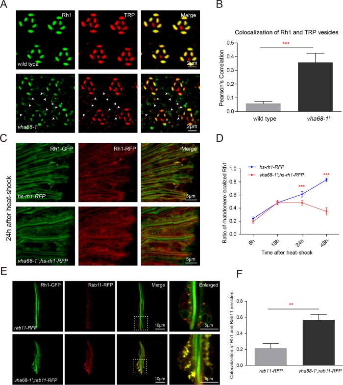
FIGURE 2:
Vha68-1 is required for Rh1 maturation. (A) Tangential resin-embedded retinal sections from ∼1-d-old wild-type and vha68-11 flies were labeled using antibodies against Rh1 (green) and TRP (red). Colocalized Rh1 and TRP vesicles are indicated by arrowheads in vha68-11 cell bodies. (B) Quantification of the Pearson’s correlation between Rh1 and TRP vesicles. Images of retinas from six different flies for each genotype were used for quantification. Error bars represent SD; ***, p < 0.001 (Student’s unpaired t test). (C) Cryostat sections of hs-rh1-RFP (ey-flp rh1-GFP;hs-rh1-RFP/+) and vha68-11;hs-rh1-RFP (ey-flp rh1-GFP;vha68-11 FRT40A/GMR-hid CL FRT40A; hs-rh1-RFP/+) labeled using RFP antibodies are shown. Flies were heat shocked for 1 h and kept in the dark for 24 h. GFP fluorescence of Rh1-GFP was directly observed to indicate rhabdomeres. (D) Quantification of rhabdomere-localized, newly synthesized Rh1-RFP at indicated time points after a 1-h heat shock. Six different compound eye sections of each sample were quantified. Error bars indicate SD; ***, p < 0.001 (Student’s unpaired t test). (E) Live confocal imaging of dissected ommatidia from rab11-RFP (rh1-GFP;ninaE-rab11-RFP/+) and vha68-11;rab11-RFP (ey-flp rh1-GFP;vha68-11 FRT40A/GMR-hid CL FRT40A;ninaE-rab11-RFP/+) flies. Images with high magnification are shown in right panels. (F) Quantification of colocalization between Rh1 and Rab11 vesicles. At least 15 ommatidia from three compound eyes of each genotype were quantified. Error bars indicate SD; **, p < 0.01 (Student’s unpaired t test).

