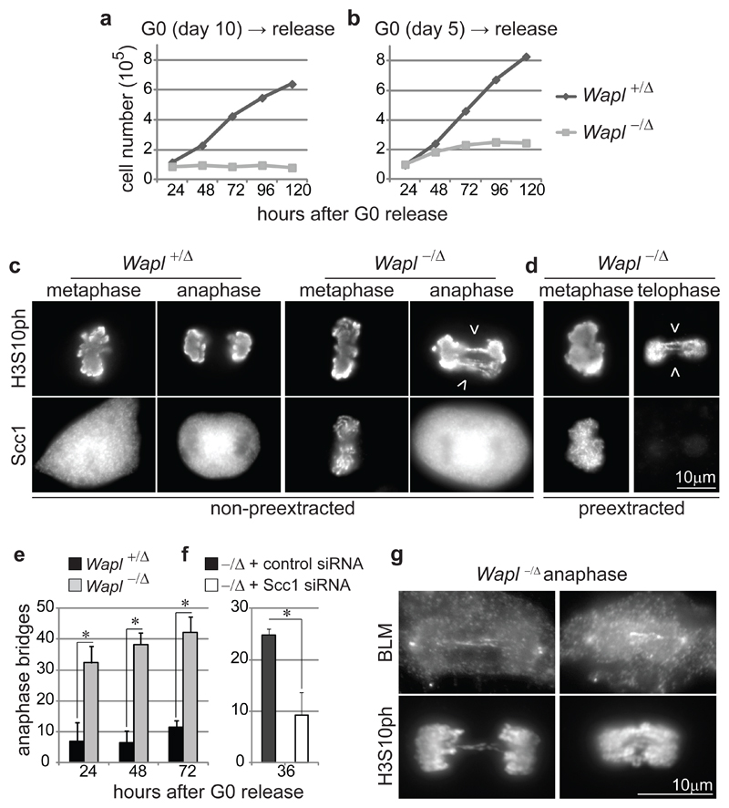
Figure 3. Wapl is essential for cell cycle progression and proper chromosome segregation.
a,b Proliferation curves of MEFs obtained as in Fig. 1a. c,d IFM images of metaphase, anaphase and telophase MEFs 72 hours after G0 release as in (b), either pre-extracted (d) or not (c) and co-stained for Scc1 and histone H3 phosphorylated on serine 10 (H3S10ph). Arrowheads indicate chromosome bridges. e, Quantification of chromosome bridge frequency in MEFs from (c, d), shown as mean and s.d. of three experiments (n≥ 100/condition; asterisk, P< 0.05, two-tailed Student’s test). f, Quantification of chromosome bridge frequency in Wapl -/Δ MEFs treated with Scc1 or control siRNAs, shown as mean and s.d. of three experiments (n≥ 156/condition; P< 0.05). g, IFM images of Wapl -/Δ anaphase MEFs 48 hours after G0 release (b) co-stained for BLM and H3S10ph.

