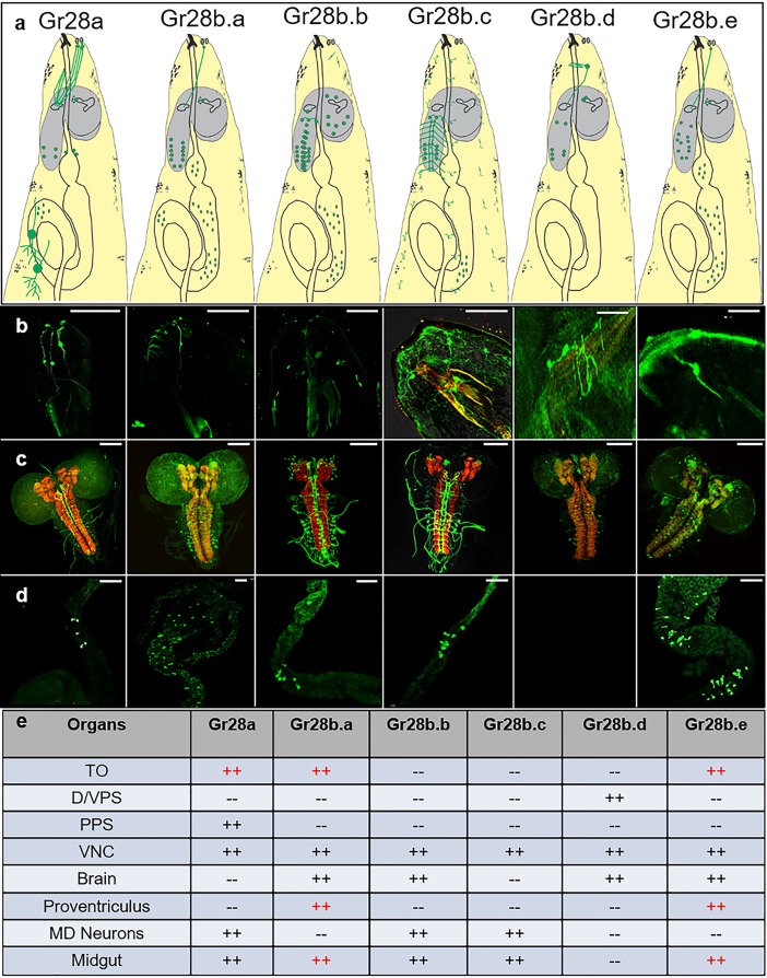
Fig 2. Expression of the 6 Gr28 genes in third-instar larvae.
(a) Graphic summary of Gr28 gene expression. Cells and neurons with their axons expressing the respective GAL4 driver are all shown in green. Brain is shown in grey, and the digestive system—including the pharynx, PV, and gut—are outlined. (b) Live GFP imaging of the larval head, showing expression of 3 genes (Gr28a, Gr28b.a, and Gr28b.e) in neurons of the TO. Gr28b.d is expressed in neurons of the DPS and VPS organs, while Gr28a is also expressed in the PPS organ. Neither Gr28a nor Gr28b.d are co-expressed with Gr43GAL4 (see S1 Fig). Number of larvae with GFP positive taste neurons/total number of larvae analyzed were 7/7 for Gr28a, 4/4 for Gr28b.a, 0/5 for Gr28b.b, 0/7 for Gr28b.c, 5/5 for Gr28b.d, and 5/5 for Gr28b.e. (c) View of the brain and parts of the ventral nerve cord, showing different degrees of expression for each of the 6 Gr28 genes. The brains were stained with anti-GFP antibody (green) and counterstained with nc82 antibody (red). Number of larvae with GFP antibody–positive staining in the brain-VNC/number of brains analyzed were 3/3 for Gr28a, 5/5 for Gr28b.a, 3/3 for Gr28b.b, 5/5 for Gr28b.c, 3/3 for Gr28b.d, and 6/6 for Gr28b.e. (d) Live GFP imaging of the PV and midgut, showing expression of all Gr28 genes with the exception of Gr28b.d. Expression of Gr28b.a and Gr28b.e is broad and includes the PV and midgut, while expression of Gr28a and Gr28b.b is defined to a smaller area of the gut only. Number of larvae with GFP-positive cells/total number of larvae analyzed were 5/5 for Gr28a, 4/4 for Gr28b.a, 3/3 for Gr28b.b, 5/5 for Gr28b.c, 0/5 for Gr28b.d, and 4/4 for Gr28b.e. (e) Summary of tissues expressing each of the 6 Gr28 genes. Genotypes were w; UAS-mCD8GFP/Gr28x-GAL4, such that x refers to indicated Gr-Gal4 driver. Scale bar is 100 μm. For live imaging (panel b and d), at least 5 larvae for each genotype were analyzed, and GFP cells in taste sensilla and the gut were observed in each case for Gr28a, Gr28ba, Gr28b.e, Gr28b.d, and Gr28b.e; for staining (panel c), at least 3 brains for each genotype were analyzed, with GFP-positive neurons observed in each case. The images are good representatives of these experiments. DPS, dorsal pharyngeal sensory; GFP, green fluorescent protein; Gr28, gustatory receptor subfamily 28; PPS, posterior pharyngeal sensory; PV, proventriculus; TO, terminal organ; VPS, ventral pharyngeal sensory.

