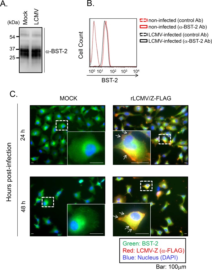
Fig 3. Effect of LCMV infection on protein expression and subcellular distribution of endogenous BST-2.
A. HeLa cells were infected with LCMV (moi = 0.01). At 48 hrs p.i, cells were lysed and BST-2 expression was analyzed by WB using a polyclonal serum to α-BST-2. B. HeLa cells were infected with LCMV (moi = 0.01). At 48 hrs p.i, cells were reacted with either control antibody (Ab) or α-BST-2 Ab conjugated with PE and then fixed. Cell surface expression of BST-2 was analyzed by FACS Calibur (BD, San Jose, CA). C. Subcellular localization of BST-2 in HeLa cells infected with rLCMV/Z-FLAG. HeLa cells were infected with rLCMV/Z-FLAG (moi = 0.01). At 24 and 48 hrs p.i., cells were fixed with 4% PFA and stained with α-FLAG antibody and α-BST-2 antibody, followed with second antibodies conjugated with Alexa 568 or Alexa 488, respectively. Nuclei were stained with DAPI. White arrows indicate LCMV Z localized at the plasma membrane without BST-2 co-localization.

