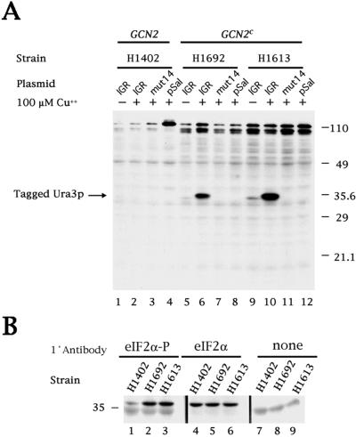
Figure 3.
Immunoblot analysis of CrPV IGR-dependent translation of tagged Ura3p and levels of eIF2α phosphorylation. (A) The transformed yeast strains are described in the legend to Fig. 1. An immunoblot was prepared and incubated with the anti-FLAG M2 monoclonal antibody to detect expression of Ura3p with a C-terminal FLAG tag. An image of the developed blot is shown. (B) Immunoblots from yeast strains H1402, H1692, and H1613 were prepared and incubated, as indicated, with anti-eIF2α polyclonal antibody, which detects only the phosphorylated form of eIF2α (eIF2α-P), with CM-217 polyclonal antibody, which detects both phosphorylated and nonphosphorylated forms of eIF2α (eIF2α) or without primary antibody (none). Images of the developed blots are shown. The 36-kDa eIF2α subunit migrates slightly above the 35-kDa marker protein.

