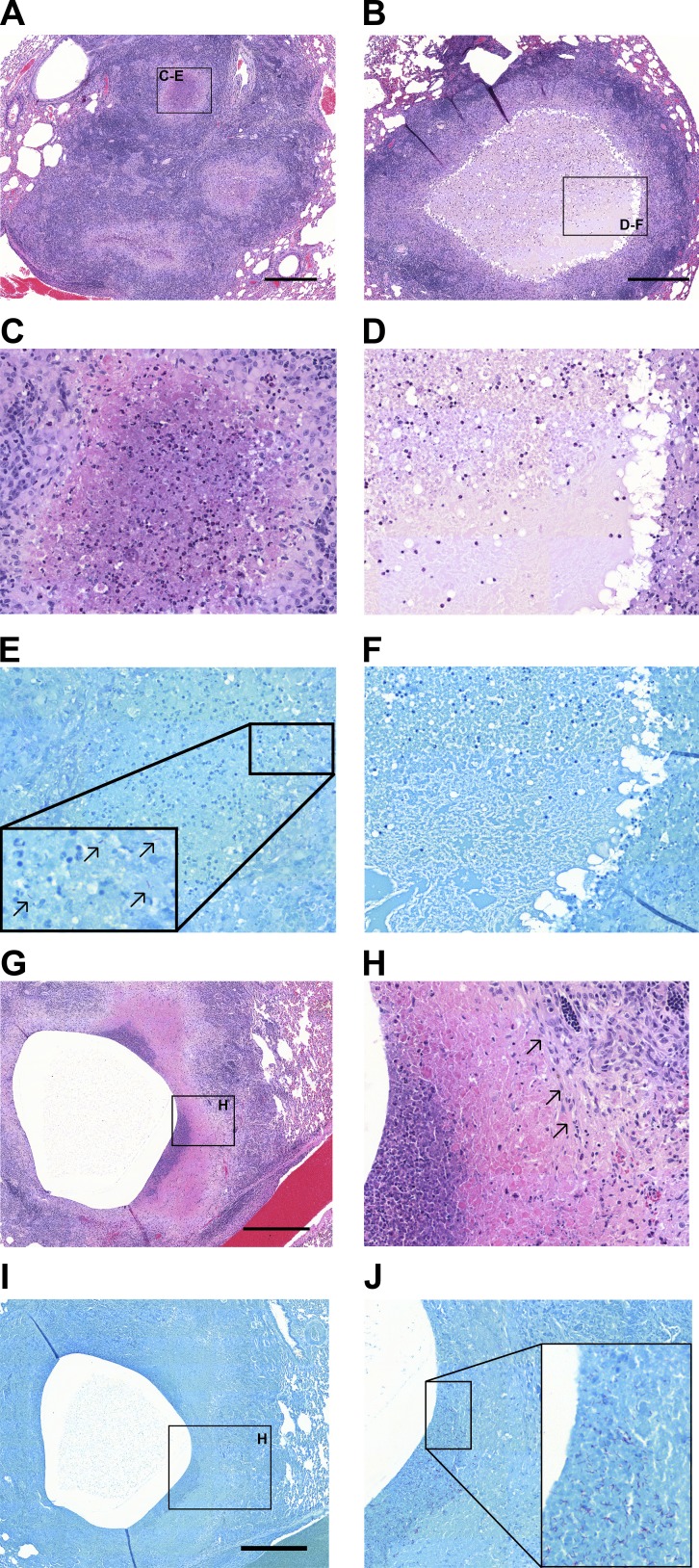
Figure 2.
Histopathological heterogeneity in rabbits with active TB and association of Mtb bacilli with neutrophils. (A) H&E staining of a cluster of small necrotizing granulomas with extensive infiltration of polymorphonuclear neutrophils in the nascent necrotic core, surrounded by a rim of epithelioid macrophages and lymphocyte-dominated inflammation at the periphery (16 wk postinfection). (B) Large necrotic granuloma with classical eosinophilic caseum in the necrotic core and low cellularity consisting of few degenerate neutrophils (16 wk postinfection). Foamy macrophages are located adjacent to the caseous necrosis. Panels C and E and panels D and F show a magnification of the regions highlighted in A and B, respectively. (C) Active neutrophil influx in the early necrotic core. (D) Caseum with limited cellular infiltrate. E and F are Ziehl-Neelsen stains of the regions shown in C and D. Black arrows in E point to Mtb bacilli found in high numbers in areas with high neutrophilic infiltration. (F) Lack of acid-fast bacilli within the paucicellular caseum. (G) H&E staining of a large cavity at 8 wk postinfection shows prior expulsion of central necrotic material and inflammatory destruction and erosion into neighboring pulmonary vein. (H) Magnification of the neutrophil-rich region highlighted in G showing the interface between low- and high-cellularity areas with high density of neutrophils. Black arrows point to an area of fibrosis at the cavity margin. (I and J) Ziehl-Neelsen stains of the regions shown in G and H, with high numbers of bacilli found in the vicinity of neutrophils. Scale bars: 500 µm.

