Figure 1.
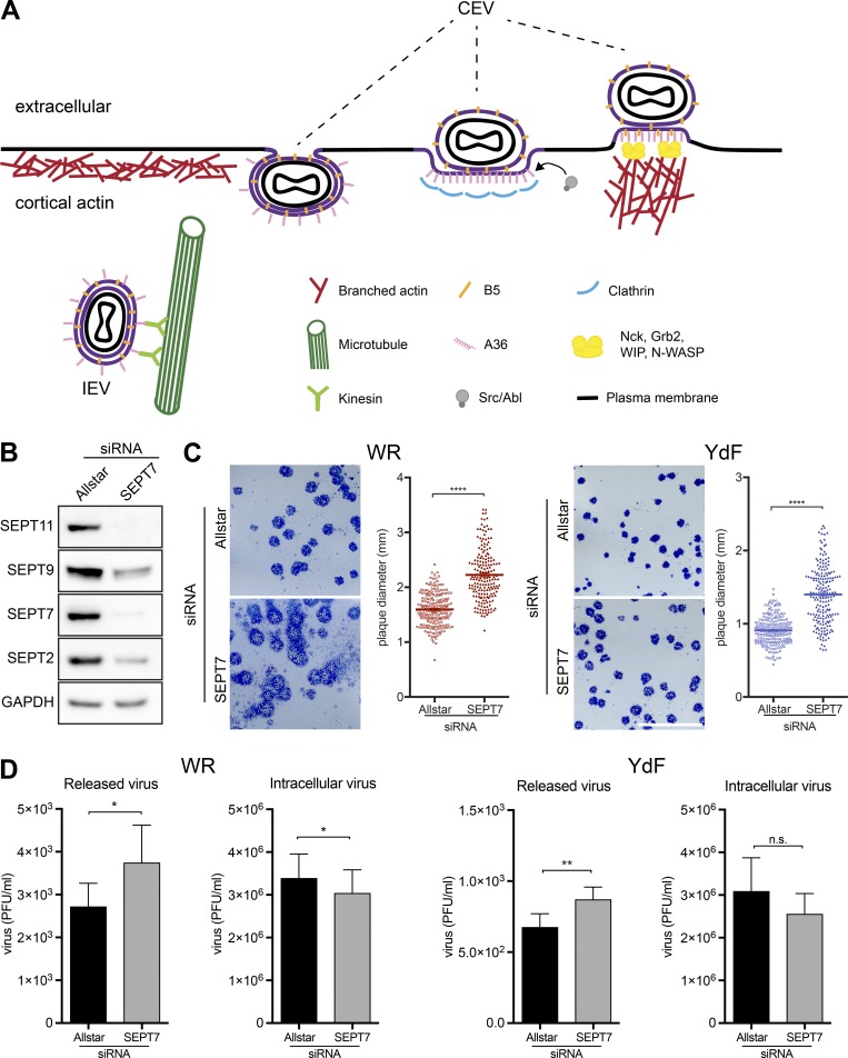
Loss of septin promotes virus release and spread. (A) Schematic depicting key events at the plasma membrane during vaccinia virus egress. A36, the actin tail nucleator phosphorylated by Src and Abl family kinases, and B5, exposed on the surface of CEV, are integral viral membrane proteins. (B) Immunoblot analysis confirms that SEPT7 siRNA treatment of A549 cells for 72 h leads to loss of SEPT7 as well as reduction in the levels of SEPT2, SEPT9, and SEPT11. (C) Images of plaques formed on A549 cell monolayers treated with control (Allstar) or SEPT7 siRNA for 72 h under liquid overlay after 3 d of infection with WR or the YdF strain of virus, which is deficient in actin tail formation. Plaque comets are seen as a diffuse spray emanating from WR plaques in liquid overlay conditions. The graphs show the quantification of plaque size (n > 190) with error bars representing the SEM from three independent experiments. (D) Quantification of virus release and total intracellular virus in A549 cells in the presence or absence of SEPT7 at 18 h after infection with WR or the YdF virus. Error bars represent SEM from three independent experiments. *, P < 0.05; **, P < 0.01; ****, P < 0.0001. Bar, 1 cm.

