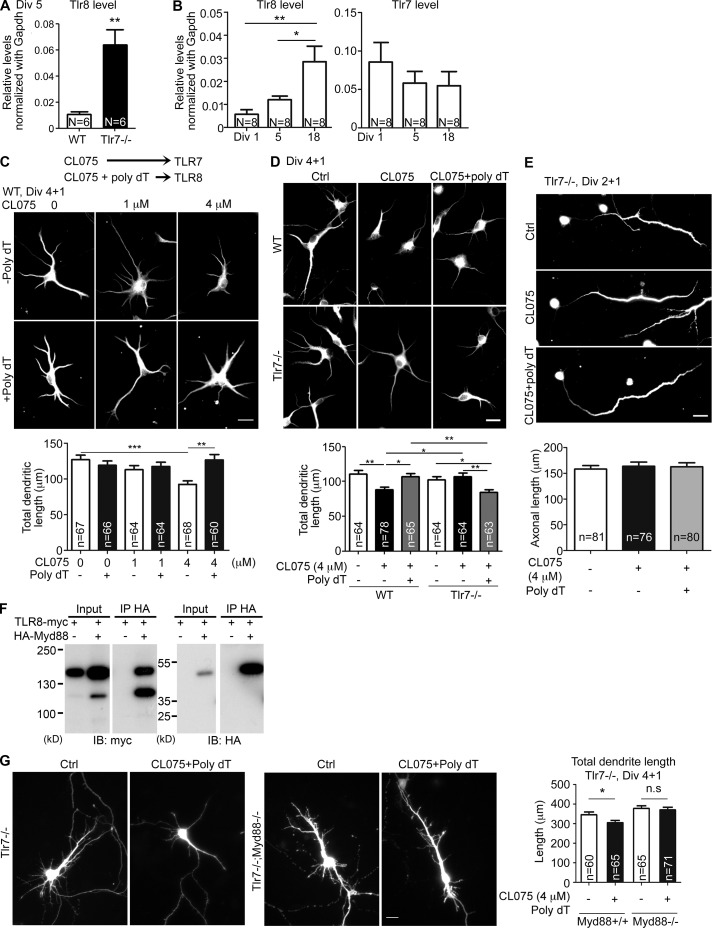
Figure 1.
Tlr8is up-regulated in Tlr7−/− neurons and restricts dendritic growth but not axonal extension of cortical and hippocampal neurons. (A and B) Up-regulated Tlr8 RNA levels in Tlr7-deficient neurons. (B) Increased Tlr8 RNA levels in mature neuronal cultures. Q-PCR was performed and normalized with internal control Gapdh. Numbers (N) of independent repeated experiments are indicated. For each experiment, three embryonic cortices and hippocampi were pooled for each set of experiments. (C and D) Different concentrations of CL075 with or without 5 µM poly dT were added to WT and Tlr7−/− cultured neurons at 4 DIV. 1 d later, neurons were harvested for immunostaining using dendritic marker MAP2 antibodies. (C) Dosage effect on WT neurons of CL075 in the presence or absence of poly dT. Based on a previous study (Gorden et al., 2006), CL075 activates TLR7 and CL075/poly dT activates TLR8. (D) Comparison of the responses of WT and Tlr7−/− neurons. (E) Tlr7−/− cultured neurons were treated with CL075 alone or mixed with poly dT at 2 DIV for 24 h and analyzed using axonal marker SMI312 antibodies. (F) Coimmunoprecipitation of Myc-tagged TLR8 and HA-tagged MYD88. The fast-migrating protein species was likely the product of TLR8 proteolysis (Lee and Barton, 2014). IB, immunoblot; IP immunoprecipitation. (G) Dendritic morphology of Tlr7−/−– and Tlr7−/−;Myd88−/−-cultured neurons at 5 DIV. Bars, 20 µm. The sample size (n) indicates the number of examined neurons, which were randomly collected blind from two (C, D, and F) or three (E) independent experiments. Data are presented as mean + SEM (error bars). *, P < 0.05; **, P < 0.01; ***, P < 0.001. Two-tailed nonparametric Mann–Whitney test (A) and one-way (B and E) and two-way (C, D, and G) ANOVA with Bonferroni’s multiple comparisons tests were used.

