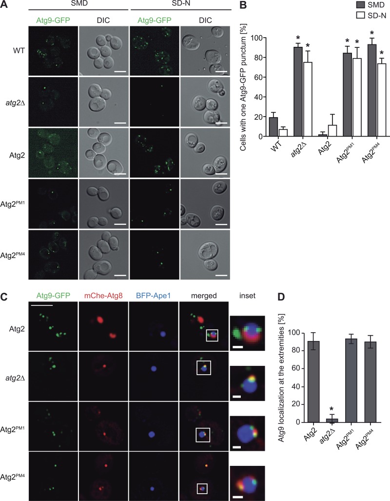
Figure 7.
Atg9 interaction with Atg2 is required for Atg9 normal subcellular distribution. (A) Localization of endogenous Atg9-GFP in WT (KTY97) or atg2Δ (SAY118) cells transformed with integrative plasmids expressing TAP-tagged versions of Atg2 (pATG2-TAP(405); RSGY003), Atg2PM1 (pATG2PM1-TAP(405); RSGY004), or Atg2PM4 (pATG2PM4-TAP(405); RSGY005) strains was analyzed. DIC, differential interference contrast. (B) Quantification of the percentage of cells displaying a single Atg9-GFP punctum in the experiment shown in A. (C) Examination of Atg9-GFP distribution on the phagophores adjacent to giant Ape1 by fluorescence microscopy. The atg2Δ mutant expressing Atg9-GFP and mCherry-Atg8 (CUY10934) was transformed with the pDP105 plasmid and analyzed as described in Materials and methods. Bars: (main images) 5 µm; (insets) 1 µm. (D) Statistical evaluation of phagophores displaying Atg9-GFP at their extremities. Graphs represent means of three experiments ± SD. Asterisks highlight significant differences with the strain carrying WT Atg2.

