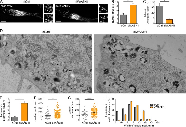
Figure 5.
The WASH complex promotes melanosomal tubules constriction and fission. (A) Live imaging frames on mCh-VAMP7 expressing MNT-1 cells treated with control or WASH1 siRNAs. Magnified areas (4×) of boxed regions show tubules (arrowheads) associated with melanosomes (arrows). (B and C) Relative number of mCh-VAMP7+ melanosomal tubules (B) detaching from melanosomes (C) during 40-s acquisition per 256-µm2 area of cells treated as in A (siCtrl, n = 4 independent experiments; siWASH1, n = 3 independent experiments). (D) High-pressure frozen control- or WASH1-depleted MNT-1 cells analyzed by 2D EM. Large tubules (arrowheads) emerge from melanosomes (arrows) upon siWASH1 treatment. (E) Percentage of melanosomes positive for tubule per n cell on EM section (siCtrl, n = 27; siWASH1, n = 21). (F and G) Mean length (F) of n tubules and width (G) of the neck (siCtrl, n = 41; siWASH1, n = 139). (H) Frequency of distribution of the width of tubules neck. Data are presented as the mean ± SEM. Bars: (A) 10 µm; (D and magnifications in A) 1 µm. ****, P < 0.0001; **, P < 0.01; *, P < 0.05 (unpaired t test).

