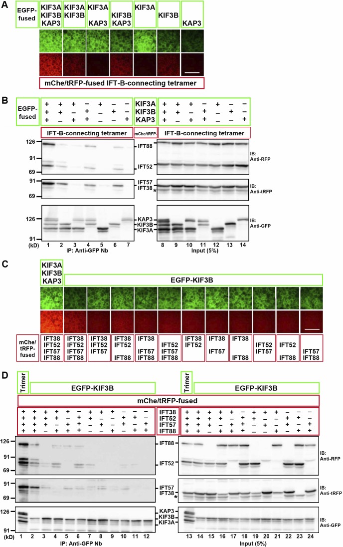
Figure 3.
KIF3B mainly contributes to the interaction with the IFT-B–connecting tetramer. (A and B) Subtractive VIP assay and immunoblotting (IB) analysis to determine subunits of kinesin-II required for its interaction with the IFT-B tetramer. (C and D) Subtractive VIP assay and immunoblotting analysis to determine subunits of the IFT-B–connecting tetramer required for its interaction with KIF3B. Details are essentially the same as described in the legend for Fig. 2 (C and D), except that exposure time for red fluorescence was 0.2 s. Bars, 100 µm. In B and D, asterisks indicate the position of mChe-IFT52, with which anti–tRFP antibody cross-reacted.

