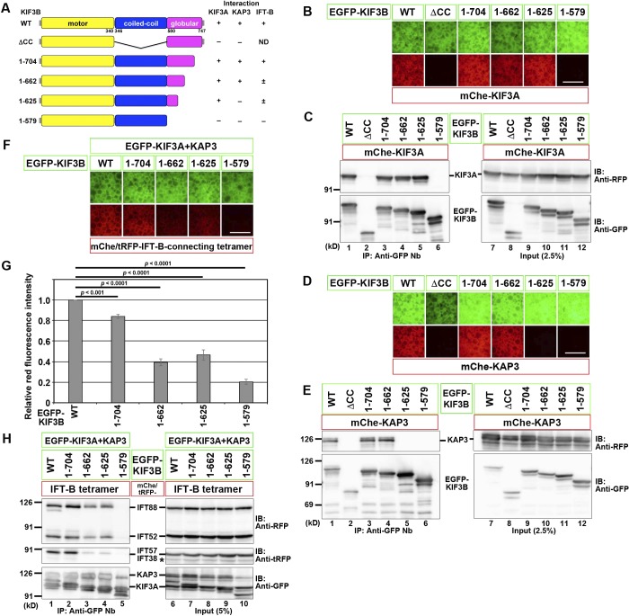
Figure 4.
Regions of the KIF3B protein responsible for its interactions with KIF3A, KAP3, and the IFT-B–connecting tetramer. (A) Schematic representation of the KIF3B constructs used and their domain organizations. On the right side, interactions of these constructs with KIF3A, KAP3, and the IFT-B–connecting tetramer are summarized. +, strong interaction; ±, weak interaction; −, no interaction; ND, not determined. (B–F and H) VIP assay (B, D, and F) and immunoblotting (IB) analysis (C, E, and H) to determine the regions of KIF3B responsible for its interaction with KIF3A (B and C), KAP3 (D and E), or the IFT-B–connecting tetramer (F and H). Bars, 100 µm. In H, an asterisk indicates the position of mChe-IFT52, with which anti–tRFP antibody cross-reacted. (G) Red fluorescence intensities in the acquired images shown in F were measured using ImageJ, and relative fluorescence intensities are expressed as bar graphs. Values are means ± SD of three independent experiments. P-values were determined by one-way ANOVA followed by Tukey’s post hoc analysis.

