Figure 2.
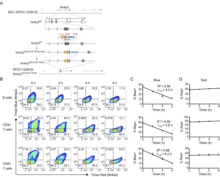
Antigen–receptor ligation induces Timer proteins in B and T cell subsets from Nr4a3-Tocky mice. (A) Construct for generating Nr4a3-Tocky BAC transgenic mice. (B) Splenocytes from Nr4a3-Tocky mice were activated for 20 h with either 10 µg/ml of soluble goat anti–mouse IgM (for CD19+ B cells) or 2 µg/ml plate bound anti-CD3 (for CD4+ and CD8+ T cells). Cells were then incubated with 100 μg/ml cycloheximide to inhibit new protein translation and the decay of blue fluorescence measured over time by flow cytometry. Shown is Timer–blue versus Timer–red fluorescence in CD19+ B cells (top), CD4+ T cells (middle), or CD8+ T cells (bottom) at the indicated time points. (C and D) Summary data of the percentage of cells blue+ (C) or red+ (D) cells in the cultures. Linear regression by Pearson’s correlation; n = 3 culture triplicates. Data are representative of two independent experiments.

