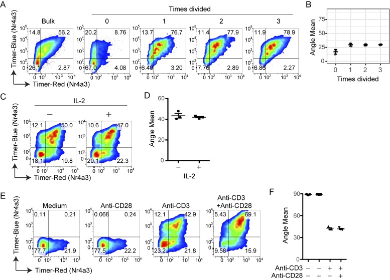
Figure 5.
Cell division, costimulation, and IL-2 signaling do not affect Timer Angle progression. (A–F) CD4+ T cells from Nr4a3-Tocky mice were labeled with a proliferation dye and activated for 72 h with anti-CD3. Cells were then analyzed based on dilution of proliferation dye and classified into number of cellular divisions. (A) Timer–blue versus Timer–red fluorescence in CD4+ T cells gated on dilution of proliferation dye. (B) Mean Timer Angle values in the cultures from A. (C and D) Splenocytes from Nr4a3-Tocky mice were stimulated on anti-CD3–coated plates in the presence or absence of 100 U/ml rhIL-2 for 20 h. Timer–blue versus Timer–red fluorescence in CD4+ T cells from cultures (C). Mean Timer-Angle in cultures (D). (E and F) Splenocytes from Nr4a3-Tocky mice were stimulated on plates coated with anti-CD28 alone, anti-CD3 alone, or anti-CD3 + anti-CD28 for 20 h. Timer–blue versus Timer–red fluorescence in CD4+ T cells from cultures (E). Mean Timer-Angle in cultures (F). n = 3 culture triplicates; error bars represent mean ± SEM. Data are representative of two independent experiments.

