. 2018 Aug 7;7:e37168. doi: 10.7554/eLife.37168

Figure 3. Compound 147 covalently modifies cellular proteins.

(A) Bar graph showing the relative activation of the ERSE.FLuc ATF6 reporter in HEK293T cells treated with 147 (10 µM), 147–20 (10 µM), or thapsigargin (Tg; 0.5 µM) for 18 hr. Error bars show SEM for three technical replicates. (B) Schematic showing the protocol for affinity purification of proteins covalently modified by 147–20. (C) Coomassie-stained SDS-PAGE of affinity purified proteins from ALMC-2 cells treated with 147 (10 µM), or 147–20 (10 µM), or the combination of the 147–20 (10 µM) and 147 (50 µM), or 147–20 (10 µM) and 147–4 (50 µM) in combination for 18 hr. Specific proteins identified by mass spectrometry of excised bands are indicated.
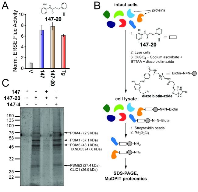
Figure 3.

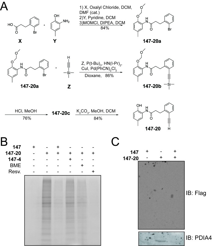
Figure 3—figure supplement 1. Compound 147 covalently modifies cellular proteins.

Figure 3—figure supplement 1.

(A) Synthetic scheme for the preparation of 147–20. (B) Coomassie-stained SDS-PAGE of affinity purified proteins from ALMC-2 cells treated with 147 (10 µM), or 147–20 (10 µM), or the combination of 147–20 (10 µM) and 147 (50 µM), or the combination of 147–20 (10 µM) and 147–4 (50 µM),or the combination of 147 (10 µM) and β-mercaptoethanol (BME; 110 μM), or the combination of 147 (10 µM) and resveratrol (10 µM) for 18 hr. (C) Immunoblot of affinity purified FLAG-tagged ATF6 from HEK293T cells stably expressing FLAG-tagged ATF6 treated with 147 (10 µM), 147–20 (10 µM), or 147–20 (10 µM) and 147 (50 µM) in combination for 18 hr.