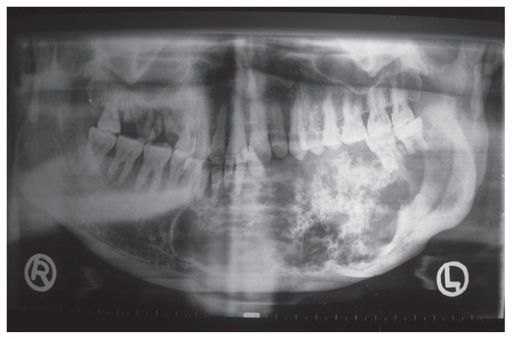
Figure 2.
Orthopantomograph showing a mixed radiolucentradiopaque multilocular lesion, extending from the right parasymphysis to the left angle region of the mandible, with welldefined, sclerotic, scalloped borders. Also notice displacement of teeth 31 and 32 and root resorption of 41, 32, 37 and 38.

