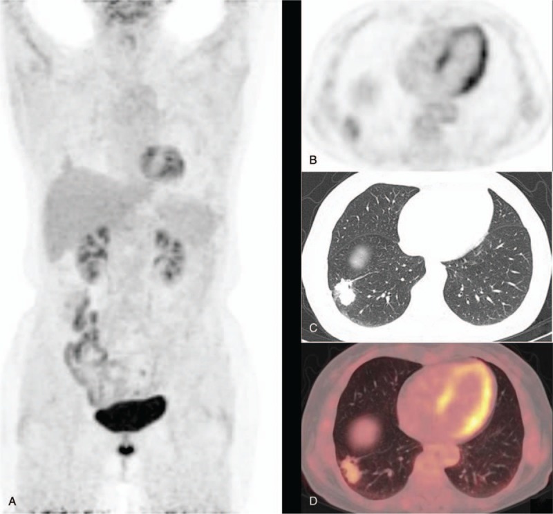
Figure 1.

A 69-year-old female patient presented with a subsolid nodule outside the basal segment of the lower lobe of the right lung. The diameter of the nodule measured about 29 mm. The SUVmax and delayed SUVmax were 2.43 and 3.12, respectively. Histopathology revealed a highly differentiated adenocarcinoma. A shows PET MIP. B shows a PET image in transverse section. C shows a chest thin layer CT image. D shows a fusion image of PET/CT. PET/CT = positron emission tomography/computed tomography, SUVmax = maximum standardized uptake value. MIP = maximum intensity projection.
