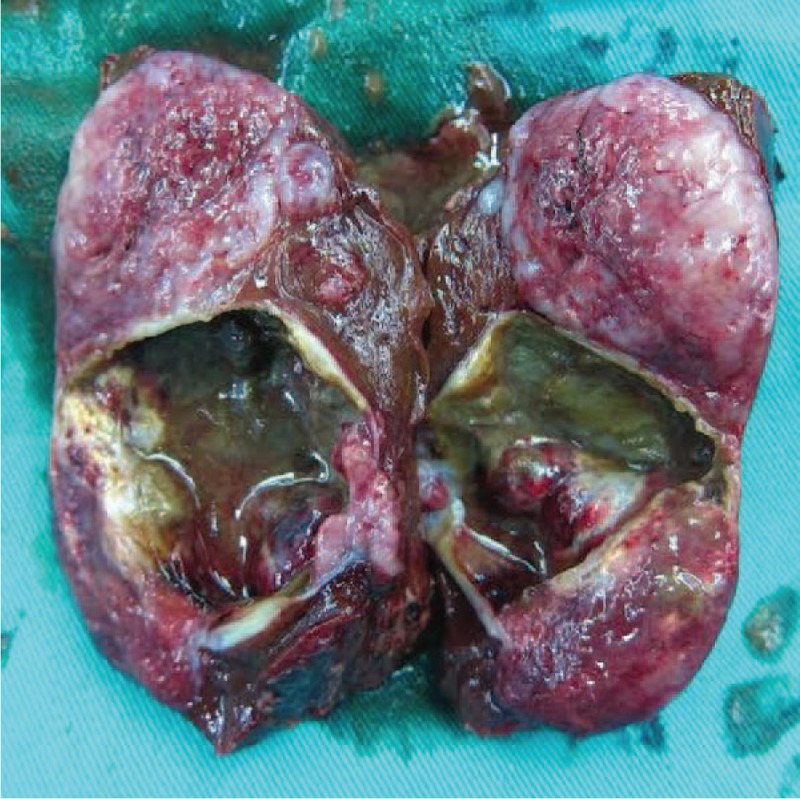
Figure 5.

The cut surface showed a yellowish-tan to gray-red solid parenchyma with focal irregular cystic spaces containing colorless serous fluid.

The cut surface showed a yellowish-tan to gray-red solid parenchyma with focal irregular cystic spaces containing colorless serous fluid.