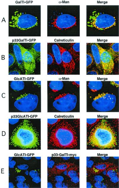
Figure 2.
Localization of chimeric enzymes involved in the formation of linkage region. Wild-type CHO cells were transiently transfected with the indicated chimeric enzymes and imaged by deconvolution microscopy (Materials and Methods). (A and C) Golgi localization of GalTI-GFP and GlcATI-GFP, respectively. (B and D) ER localization of p33-GalTI-GFP and p33-GlcATI-GFP, respectively. (E) Coexpression of GlcATI-GFP (green) and p33-GalTI-Myc (red). α-Man, α-mannosidase II, the medial Golgi marker; Calreticulin, ER marker; Merge, colocalization (yellow) of the chimeric enzymes with each other or the markers.

