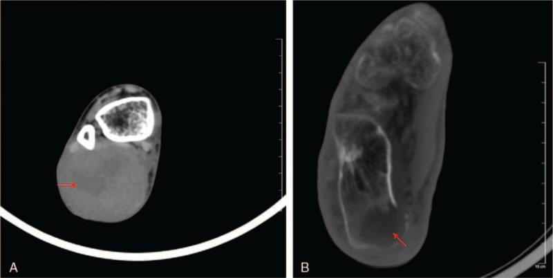
Figure 2.

(A) CT axial view—soft tissue window showing a heterogeneous density within a well-defined mass in the posterior compartment of the right ankle (red arrow). (B) Axial view—bone window demonstrating a hypodense osteolytic lesion in the calcaneal bone (red arrow). CT = computed tomography.
