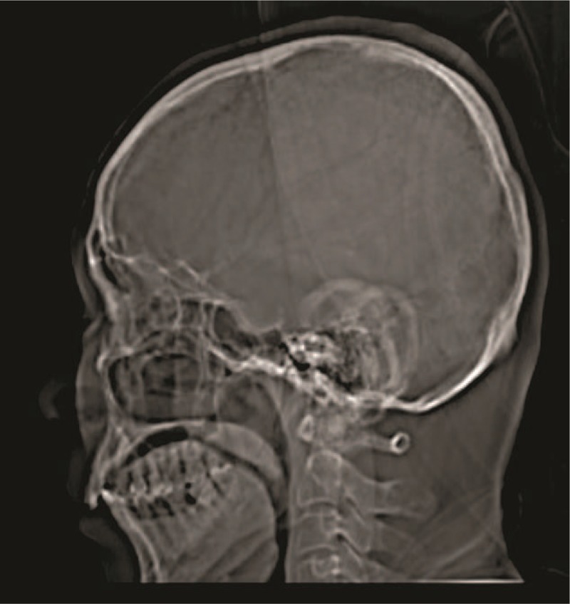
Figure 2.

The computed tomographic image of 35 years old patient, who had pituitary adenomas. 0.6 cm slice thickness, 80 kV voltage, 75 μA current, 20 s/scan, and 360o rotation, 5 mSv radiation dose, display field of view: 51.2 × 61.5 cm, and zoom: 144%.
