Figure 5.

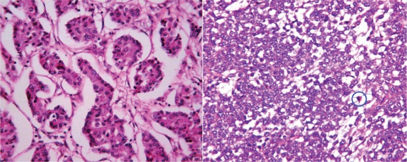
HE×400: (A) Microscopic findings for a PHNEN (G3), revealing tumor cells aligned in a nest-like structure and surrounded by cuboidal cells. Cancer nests were encased by blood sinusoids. (B) Mitosis in the examined circle.

HE×400: (A) Microscopic findings for a PHNEN (G3), revealing tumor cells aligned in a nest-like structure and surrounded by cuboidal cells. Cancer nests were encased by blood sinusoids. (B) Mitosis in the examined circle.