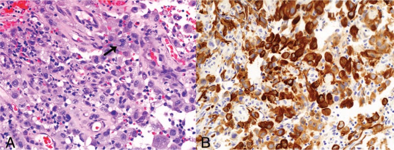
Figure 2.

Biopsy of tumor at initial presentation. (A) Sheets of infiltrating pleomorphic tumor cells, with atypical mitoses (arrow) (hematoxylin and eosin stain, original magnification ×200). (B) The tumor cells demonstrate strong diffuse positive staining with AE1/3 immunostain (original magnification ×200).
