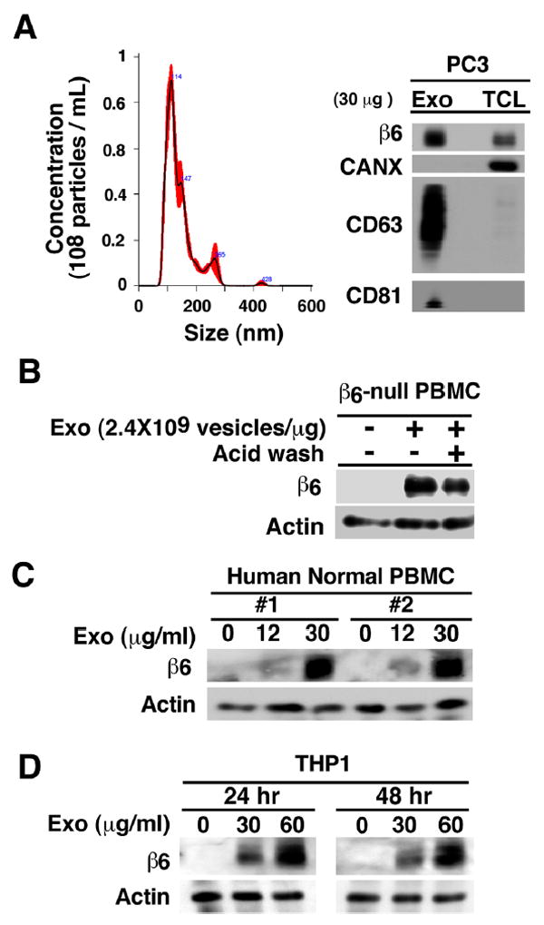
Fig. 1. Exosomal αvβ6 is transferred from prostate cancer cells to PBMC.

(A), Left, nanoparticle size distribution analysis of PC3 exosomes (Exo) by NTA. Right, IB analysis of β6 integrin, exosomal markers CD63, CD81 and calnexin (CANX) in lysates of PC3 Exo and cells (TCL). (B), PBMC (3.0×10⁵ cells) derived from β6-null mice (pool of 3 mice) were incubated with PC3 Exo (30 μg/mL at a concentration of 2.4×10⁹ vesicles/μg) for 24 hours. The cells were washed with acid wash buffer twice followed by IB analysis of cell lysates for expression of β6 integrin and actin (loading control). (C), PBMC (3.0×10⁵ cells) from two different human healthy donors were incubated with indicated PC3 Exo concentrations (0, 12, 30 μg/mL at a concentration of 2.4×10⁹ vesicles/μg) for 24 hours and cell lysates were analyzed by IB for expression of β6 integrin and actin (loading control). (D), THP1 cells (3.0×10⁵ cells) were incubated with the indicated PC3 Exo concentrations (0, 30, 60 μg/mL at a concentration of 2.4×10⁹ vesicles/μg) for 24 and 48 hours and analyzed by IB for expression of β6 integrin and actin (loading control).
