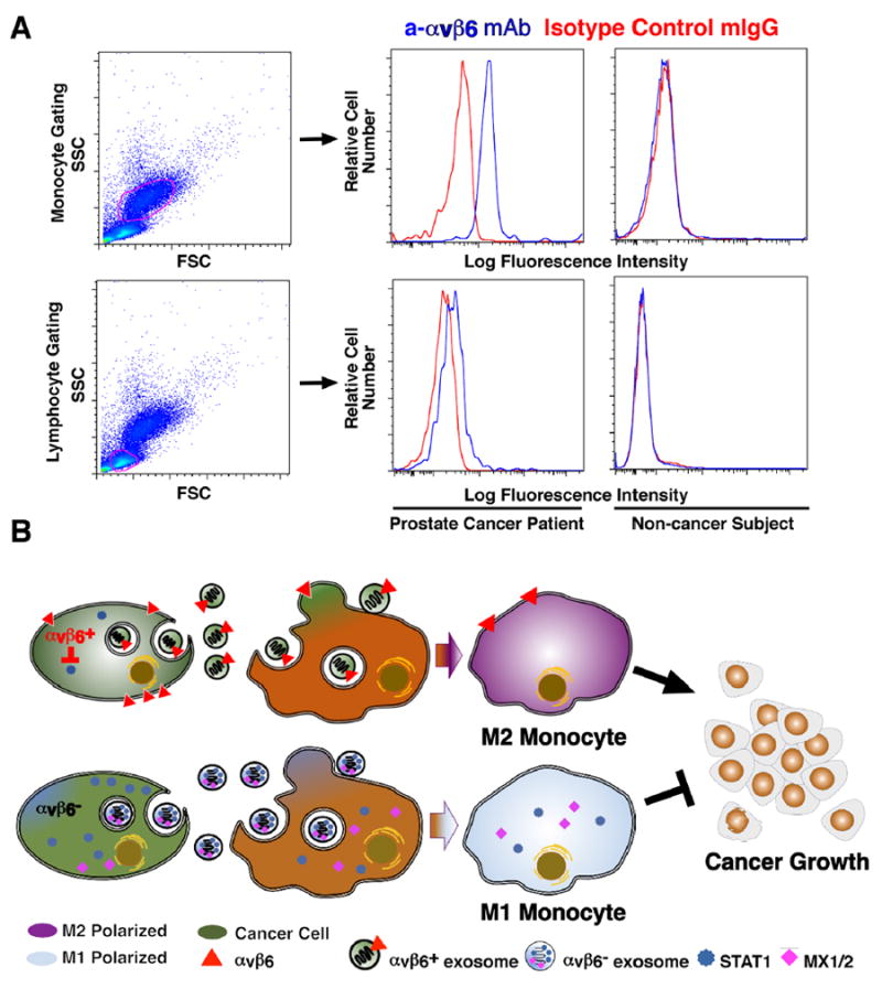
Fig 7. αvβ6 Integrin is expressed in PBMC from prostate cancer patients.

(A), Flow cytometric analysis of αvβ6 expression in PBMC from prostate cancer patients and healthy subjects. Left, monocytes and lymphocytes are gated by SSC and FSC. Right, FACS analysis of αvβ6 cell surface expression in monocytes and lymphocytes from healthy subjects and prostate cancer patients respectively, utilizing 6.3G9 monoclonal antibody to αvβ6 and mouse IgG, as isotype control. Representative data are shown. (B), The schematic diagram shows that transfer of exosomal αvβ6 integrin from prostate cancer cells to monocytes results in down-regulation of STAT1 and MX1/2 levels and increased M2 polarization of monocytes, and has a pro-tumorigenic effect, whereas transfer of αvβ6 integrin–negative exosomes results in increased M1 polarization of monocytes, and inhibits cancer growth
