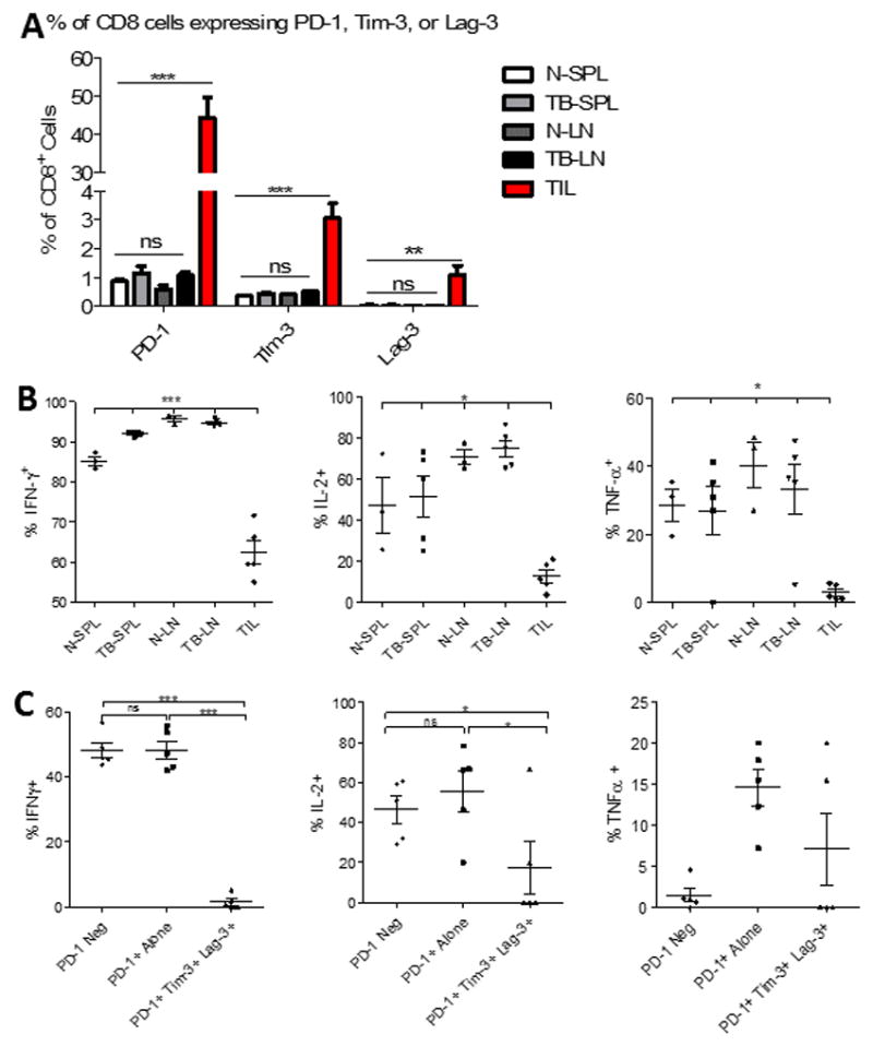
Figure 3. TIL isolated from murine gliomas demonstrate impaired function.

A. The percentage of CD8+ T cells expressing either PD-1, Tim-3, or Lag-3 alone was compared across immunologic compartments. T cells were isolated from the spleens (SPL) and cervical lymph nodes (LN) from either CT2A tumor-bearing (TB) (n=5) or naïve (N) (n=3) mice, as well as from tumors of TB mice (TIL). B. The capacity for CD8+ T cells isolated from the same sites to express IFN-γ, IL-2, and TNF-α, upon stimulation with PMA/ionomycin was compared. C. Boolean gating was employed to determine percentage of cells producing cytokines among TIL not expressing PD-1, PD-1 single positive, or PD-1/TIM-3/LAG-3 triple positive cells. Statistical significance was assessed via unpaired t-test between control and tumor-bearing samples. *p<0.05; **p<0.01; ***p<0.0001 throughout figure.
