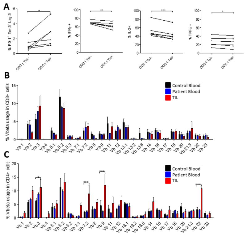
Figure 5. T cell exhaustion arises preferentially amongst tumor-specific T cells.

A. 5 mice were implanted with SMA-560 tumors IC. Tumors were harvested when mice were moribund and TIL isolated. TIL were stimulated with the ODC-1 peptide for 6 hours, stained with the ODC-1 tetramer conjugated to APC, several immune checkpoints and intracellular stain for cytokines was performed. Significance was assessed via paired t-test between tetramer positive and negative cells. Vbeta analysis was performed on CD8+ (B) and CD4+ (C) T cells isolated from human GBM TIL (n=5), patient blood (n=5), or control blood (n=5). Significance was assessed using a two-way ANOVA to assess for interaction between Vb and sample type, followed by Bonferroni post-tests between patient and control samples. *p<0.05; **p<0.01; ***p<0.001.
