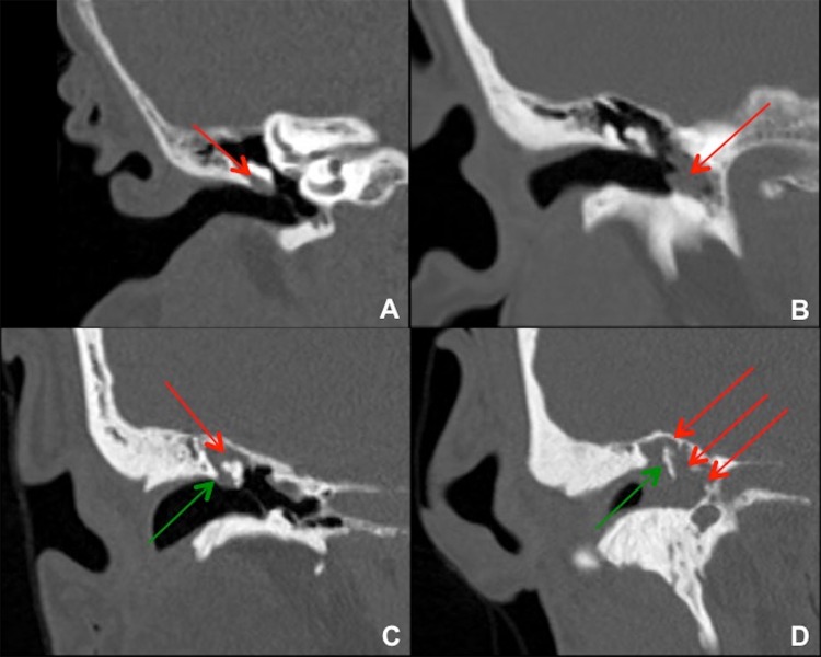
Fig. 1.
Coronal images from temporal bone CTs in four different patients with right cholesteatoma. a Small cholesteatoma in Prussak’s space (red arrow) without bony erosion. This is a common site for pars flaccida retraction and acquired cholesteatoma formation. b Cholesteatoma in the left mesotympanum to hypotympanum (red arrow), which is a less common site. c Cholesteatoma in the right epitympanum (red arrow) with blunting or erosion of the right scutum (green arrow). This lesion probably started in Prussak’s space adjacent to the bony scutum. d Large cholesteatoma in the right epitympanum, mesotympanum, and hypotympanum (red arrows), with bony erosion of the scutum and malleus/ossicles (green arrow)

