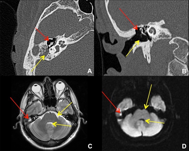
Fig. 2.
Temporal bone CT and brain MRI in 41-year-old male after transcanal endoscopic resection of a right epitympanic cholesteatoma. a Axial CT shows the resection cavity in the right Prussak’s space (red arrow), with residual cholesteatoma in the right mastoid antrum (yellow arrow). b Coronal CT shows the resection cavity at the right lateral epitympanum (red arrow) plus the surgical approach for a transcanal atticotomy (yellow arrow). An alternative approach would be via the mastoid antrum (mastoidectomy). c Axial T2-weighted MRI shows the small residual cholesteatoma (red arrow) to be of similar intensity with fluid, e.g. prepontine cistern and fourth ventricle (yellow arrows). d Axial diffusion-weighted MRI shows “restricted diffusion” in the cholesteatoma (red arrow), much brighter than free fluid (yellow arrows)

