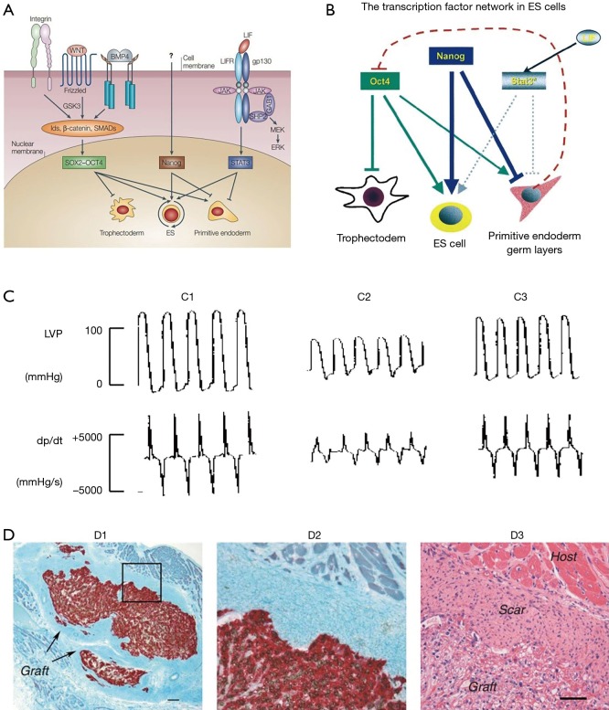
Figure 2.
Stem cells and heart regeneration. (A) Signal pathways involved in maintaining mouse ESC pluripotency. (B) Model of the transcription factor network in embryonic stem (ES) cells. The arrow indicates the promoting effect and the horizontal line means the blocking effect (21). (C) Embryonic stem cell transplantation improved left ventricular (LV) function (24). (C1) Sham-operated rats; (C2) postinfarcted rats injected with cell-free medium; (C3) postinfarcted rats transplanted with embryonic stem cells. (D) Human embryonic stem cells enhance function of infracted rat hearts (10). The bright field microscopic images were acquired from recipient hearts 4 weeks post-transplantation. (D1) The human pan-centromeric in situ hybridization (brown chromagen) and β-myosin-positive cardiomyocytes (red) indicated the formation of a large cell graft within infarct scar tissue. Scale bar =100 µm. (D2) High magnification of boxed part in D1. (D3) Hematoxylin and eosin stain showed the graft cells have a vacuolated appearance because of the glycogen. Scale bar =50 µm. [(A) reprinted with permission of reference (20), (B) reprinted with permission of reference (21), (C) reprinted with permission of reference (24) and (D) reprinted with permission of reference (10)]. LVP, left ventricular potential; dP/dt, peak LV systolic pressure.

