Figure 6.
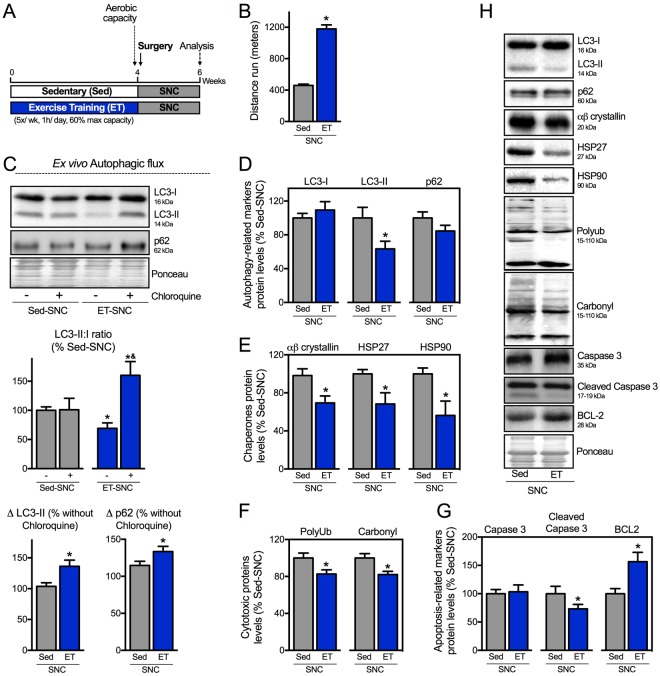
Exercise training activates autophagy and improves proteostasis in neurogenic myopathy. (A) Schematic panel of Study Design 3: rats were randomly assigned into sedentary (Sed) and exercise training (ET) groups. ET rats were submitted to running on a treadmill over 4 weeks, 5 days/week, 60 minutes per day at 60% of maximal aerobic capacity. After this period rats were submitted to SNC, and 14 days after surgery skeletal muscle morphological, functional and biochemical parameters were measured in Sed-SNC and ET-SNC rats. (B) Aerobic capacity evaluated by total distance run after protocol (week 4). (C) Ex vivo autophagic flux: LC3 and p62 protein levels and representative images of plantaris muscle from Sed-SNC and ET-SNC rats treated ex vivo with saline (−) or chloroquine (100 μg.mL−1) (+) for 4 hours. Protein levels of (D) autophagy-related markers (LC3-I, LC3-II and p62), (E) chaperones (αβ-crystallin, HSP27 and HSP90), (F) cytotoxic proteins (polyubiquitinated and protein carbonyls) and (G) apoptosis-related markers (caspase 3, cleaved caspase 3 and BCL-2), and (H) representative images of plantaris muscle from Sed-SNC and ET-SNC rats. The corresponding ponceau stain was used to verify equal loading of proteins and values are expressed as a percentage of the Sed-SNC group and as a percentage of the respective group without chloroquine (autophagic flux). Data are presented as mean ± SEM. *p < 0.05 vs. Sed-SNC; n = 8–12 animals.

