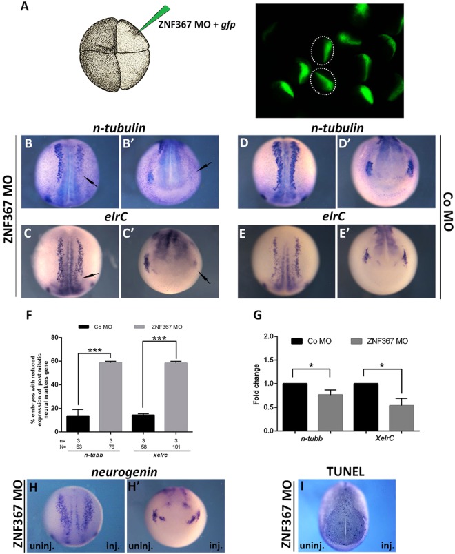
Figure 3.
Loss of znf367 function interferes with the expression of neuronal differentiation markers. (A) Embryos injected with gfp (250 pg) and either ZNF367-MO or Control morpholino (Co-MO) (9 ng) at one dorsal blastomere at the four-cells stage showed fluorescence only in the neural plate at neurula stage (st. 18). (B–E’) mRNA distribution of N-tubulin and elrC in znf367 morphants and controls. (B,C) dorsal view and (B’,C’) frontal view of neurula morphants showing a clear loss of differentiation markers expression in primary neurons (arrows). (D,E) dorsal view and (D’,E’) frontal view of neurula embryos injected with Co-MO. (F) Quantification of the data in A and B. (G) qRT-PCR analysis. The levels of expression for the indicated mRNAs were evaluated for Co-MO or ZNF367-MO populations by qRT-PCR, and normalized to that of the housekeeping gene, glyceraldehyde 3-phosphate dehydrogenase (gapdh) expression. The mean of the Control-MO was set to 1. For each gene, three independent RNA samples from morphants and controls were analysed. (H–H’) mRNA distribution of neurogenin in znf367 morphants. (I) TUNEL staining. ZNF367-MO injection did not lead to an increase of TUNEL positive cells compared to the un-injected side. Abbreviations: n, number of independent experiments; N, number of evaluate embryos in total; Error bars indicate standard error of the means (s.e.m); *p ≤ 0,05; ***p ≤ 0,001. P-value were calculated by Student’s t-test.

