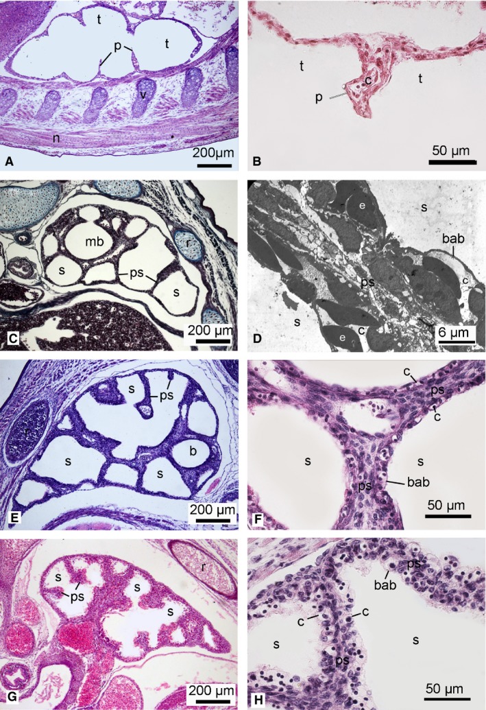
Figure 7.

Histological sections of the lung in the neonate Dasyurus viverrinus (A,B), Monodelphis domestica (C), Trichosurus vulpecula (E,F) and Perameles nasuta (G,H). Higher magnification micrographs show the septa between airspaces: (B,F,H) histological sections; (D) TEM micrograph of M. domestica (from the author's PhD work). The lung of the newborn D. viverrinus consisted of poorly vascularized airspaces (tubules). No double capillary septa and onlys a few portions of the blood–air barrier were present, thus showing characteristics of the canalicular stage (G1). The lungs of M. domestica, T. vulpecula and P. nasuta were at the early saccular stage. The large smooth airspaces were separated by thick primary septa with a double capillary network and a blood–air barrier was present (G2). b, bronchus; bab, blood‐air‐barrier; c, capillary; e, erythrocyte; mb, main bronchus; n, spinal cord; p, partitions; ps, primary septum; r, rib; s, sacculus; t, tubulus; v, cartilage of vertebra. Magnification is indicated by the scale bar.
