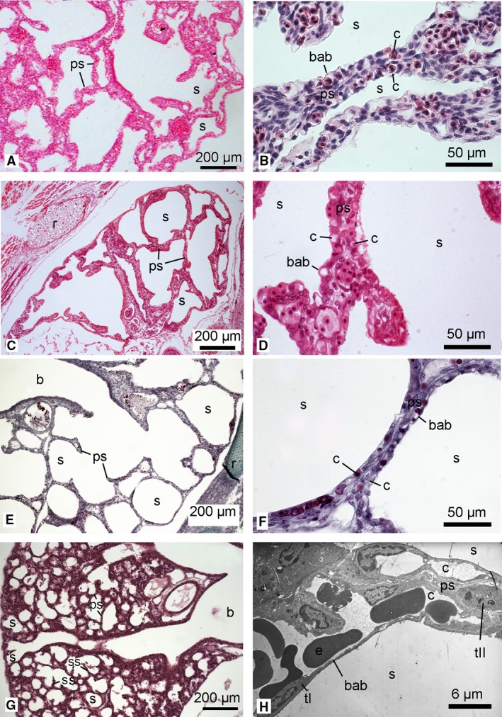
Figure 8.

Histological sections of the lung in the neonate Phascolarctos cinereus (A,B), Petrogale penicillata (C,D), O. anatinus (E,F) and Mesocricetus auratus (G). Higher magnification micrographs show the septa between airspaces: (B,D,F) histological sections; (H) TEM micrograph of M. auratus (from the author's PhD work). The lungs of P. cinereus, P. penicillata and O. anatinus were at the saccular stage, characterized by large terminal saccules separated by thick primary septa with a double capillary network. In the lungs of neonate P. cinereus and P. penicillata, numerous septal crests indicated a further proliferation of the lung parenchyma, resulting in more irregular shapes of the terminal saccules (G3). In the altricial placental neonate M. auratus, the bronchial tubes led to the periphery of the lung and terminated in numerous small saccules which were separated by a thin primary double capillary septum. A well developed blood–air barrier was present (8H). b, bronchus; bab, blood‐air‐barrier; c, capillary; e, erythrocyte; ps, primary septum; r, rib; s, sacculus; tI, type I pneumocytes; tII, type II pneumocytes. Magnification is indicated by the scale bar.
