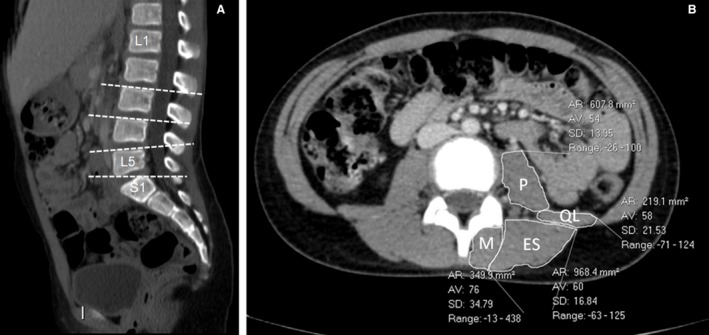
Figure 1.

(A) Sagittal reconstructed CT image of a 9‐year‐old girl showing the levels of paraspinal CSA measurements (L3, L4, L5, S1). (B) Cross‐section image at the level of L4 endplate of a 12‐year‐old boy showing the measurements of paraspinal muscles.
