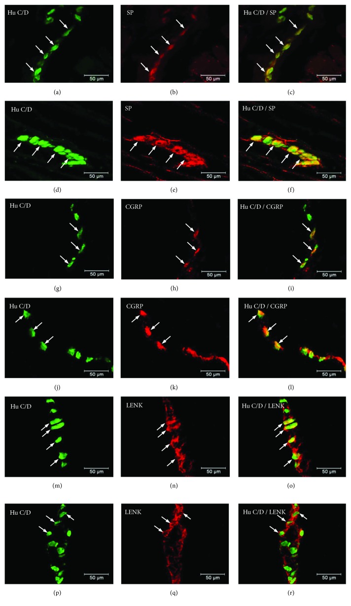
Figure 5.
Submucosal ganglion of the porcine stomach immunoreactive to SP, CGRP, and L-ENK. Submucosal ganglion of the porcine corpus under physiological condition (a–c) and during experimentally inducted diabetes (d–f) immunoreactive to SP; submucosal ganglion of the porcine corpus under physiological condition (g–i) and during experimentally inducted diabetes (j–l) immunoreactive to CGRP; and submucosal ganglion of the porcine corpus under physiological condition (m–o) and during experimentally inducted diabetes (p–r) immunoreactive to L-ENK, immunostained for Hu C/D (green/arrows) and SP,CGRP, and L-ENK (red/arrows). The right column of the pictures shows the overlap of both stainings. Colocalization of both antigens in the studied cell bodies is indicated with arrows.

