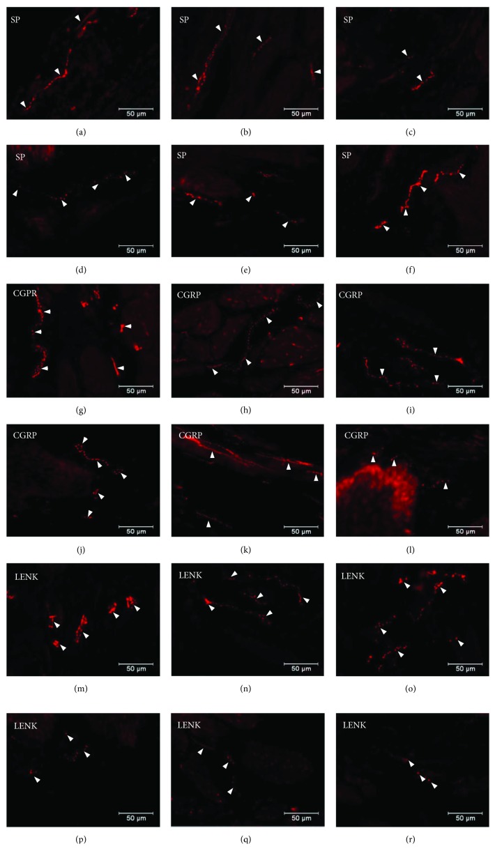
Figure 7.
Distribution pattern of nerve fibres (arrow) immunoreactive to SP, CGRP, and L-ENK within the submucosal muscle layer. Distribution pattern of nerve fibres (arrows) within the submucosal muscle layer immunoreactive to SP under physiological condition: antrum (a), corpus (b), and pylorus (c) and under experimentally inducted diabetes: antrum (d), corpus (e), and pylorus (f); for CGRP under physiological condition: antrum (g), corpus (e), and pylorus (f) and under experimentally inducted diabetes: antrum (j), corpus (k), and pylorus (l); and for L-ENK under physiological condition: antrum (m), corpus (n), and pylorus (o) and under experimentally inducted diabetes: antrum (p), corpus (q), and pylorus (r).

