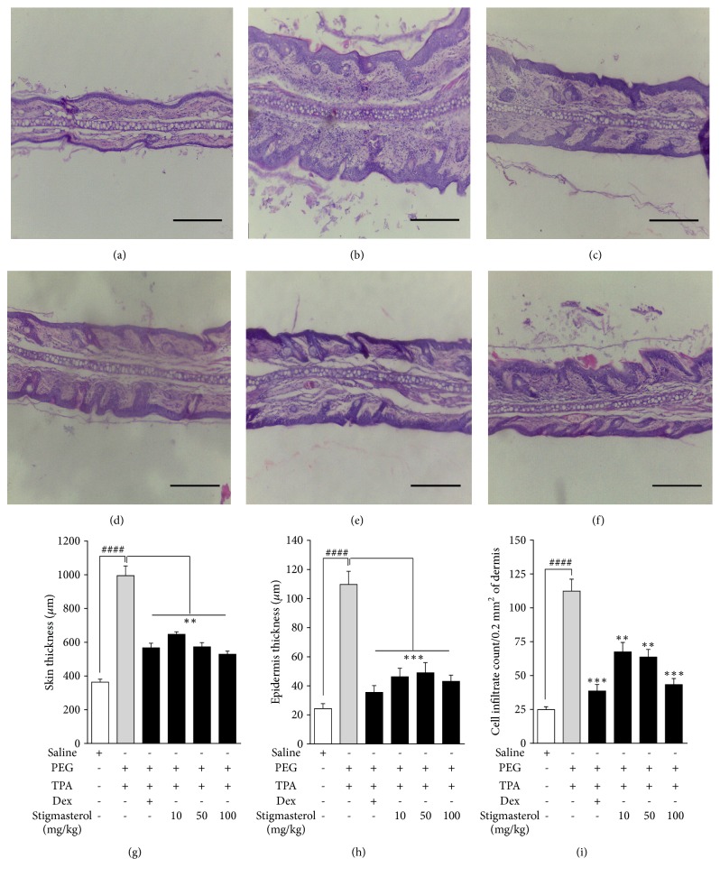
Figure 7.
Effect of stigmasterol on TPA-induced dermatitis. Rats received either normal saline (5 ml/kg), polyethylene glycol, PEG (5 ml/kg), dexamethasone, Dex (3 mg/kg), or stigmasterol (10, 50, and 100 mg/kg). Test rats received a topical application of 20 μg TPA dissolved in acetone on each ear daily for 3 days while naïve rats were challenged with acetone only. 5 h after the last TPA or acetone challenge rats were sacrificed and ears excised. 3 μm thickness of skin sections was stained with H & E and observed for histopathological changes in naïve (a), polyethylene glycol, PEG (b), dexamethasone (c), and 10-100 mg/kg stigmasterol-treated animals (d–f) and parameters of skin damage quantified (g–i). Data is expressed as mean skin thickness (μm) (n = 12) ± SEM, mean epidermis thickness (μm) (n = 12) ± SEM, and mean cell infiltrate per field (n = 12) ± SEM. ∗∗∗P < 0.001; ∗∗P < 0.01 as compared to PEG-treated TPA-challenged control. ###P < 0.0001 as compared to saline-treated naïve control using one-way ANOVA followed by Dunnett's post hoc test. Micron bar represents 500 μm.

