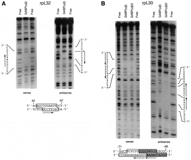
Figure 4.

DNase I footprint analysis of rpL32 and rpL30 GABP sites. Purified recombinant GABP subunits were incubated with a DNA fragment labeled at the sense or antisense strand. Free DNA and protein-bound DNA were partially digested with DNase I and subjected to electrophoresis on a 4% polyacrylamide gel in 0.5× TBE. DNA was recovered by electroelution and subjected to electrophoresis on a 8% polyacrylamide gel containing 7M urea. The positions of sequence markers are indicated at the right and left of the panels. A. Fragment corresponding to the −126 to −13 region of rpL32. B. Fragment corresponding to the −125 to −6 region of rpL30. At the bottom of the figures, the sequences of the protected regions are delineated by boxes. The stippled portion of the rpL30 box was protected in both the αβ1 and (αβ1)2 complexes; the nonstippled portion was protected only in the (αβ1)2 complex.
