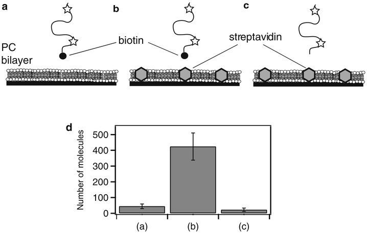
Fig. 3.
Determining the extent of nonspecific binding to a passivated surface. Data is shown for streptavidin islands (Subheading 3.1.2) but the same method can be used to test any surface preparation. (a) As a control, biotinylated, fluorescently labeled protein was applied to a deposited lipid bilayer containing no streptavidin. After 5 min incubation, the proteins were rinsed out and the extent of binding was assessed. (b) The same protein solution in (a) applied to a streptavidin island surface and rinsed. (c) As an additional control, the same protein was expressed, purified, and labeled without biotinylation and applied to the streptavidin island surface. (d) To assess the degree of binding, the number of molecules retained on the surface was counted. Shown are the average number of molecules per field of view for the experiments shown in (a)–(c), respectively. The degree of nonspecific binding can be assessed by comparing retention of the protein to the streptavidin islands to the surface without streptavidin and/or protein lacking biotin (see Note 9).

