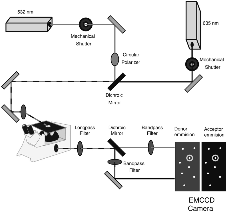
Fig. 4.
Schematic of a prism-based total internal reflection microscope for single-molecule spectroscopy. A circularly polarized 532-nm laser and a linear polarized 635-nm laser are combined using a dichroic mirror and routed to a quartz prism on the microscope stage. Mechanical shutters control the excitation wavelength. The fluorescence emission is split by color into two images using a dichroic mirror, which are passed through additional optical elements to isolate donor and acceptor signals. The two replicate images are collected by an EMCCD camera and are processed using MATLAB to identify single molecules containing a donor and acceptor fluorophore.

