Fig. 7.
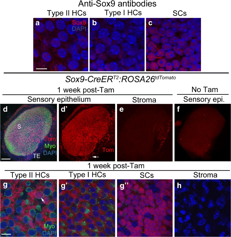
Sox9-CreERT2 mice have inducible Cre activity in supporting cells. a-c Confocal images showing immunolabeling for Sox9 (red) and nuclei (DAPI, blue) in slices focused on the nuclei of type II hair cells (HCs) (a), type I hair cells (HCs) (b), and supporting cells (SCs) (c) of the utricular macula. d-eConfocal images of the surface of utricle from Sox9-CreERT2:ROSA26tdTomato mice injected with tamoxifen, focused on either the sensory epithelium (d,d') or stroma (e). d and d' show the same view, with labeling for tdTomato (Tom, red), myosin VIIa (Myo, green), and DAPI (blue) shown in d and Tom only shown in d'. S = approximate position of the striola, TE = transitional epithelium. Arrow in d' points to labeled cells in the TE. eshows Tom labeling in the stroma. f Utricle from a Sox9-CreERT2:ROSA26tdTomato mouse that did not receive tamoxifen, focused on the macula. g-h Confocal slices through the lateral extrastriolar region, with slices through the type II hair cell (HC) nuclear layer (g), the type I hair cell (HC) nuclear layer (g'), the supporting cell (SC) nuclear layer (g"), or the stroma (h). Arrow in g indicates a Tomato-positive type II HC. Scale bar in a = 5 μm and applies to a-c. Scale bar in d = 150 μm and applies to d-f. Scale bar in g = 5 μm and applies to g-h

