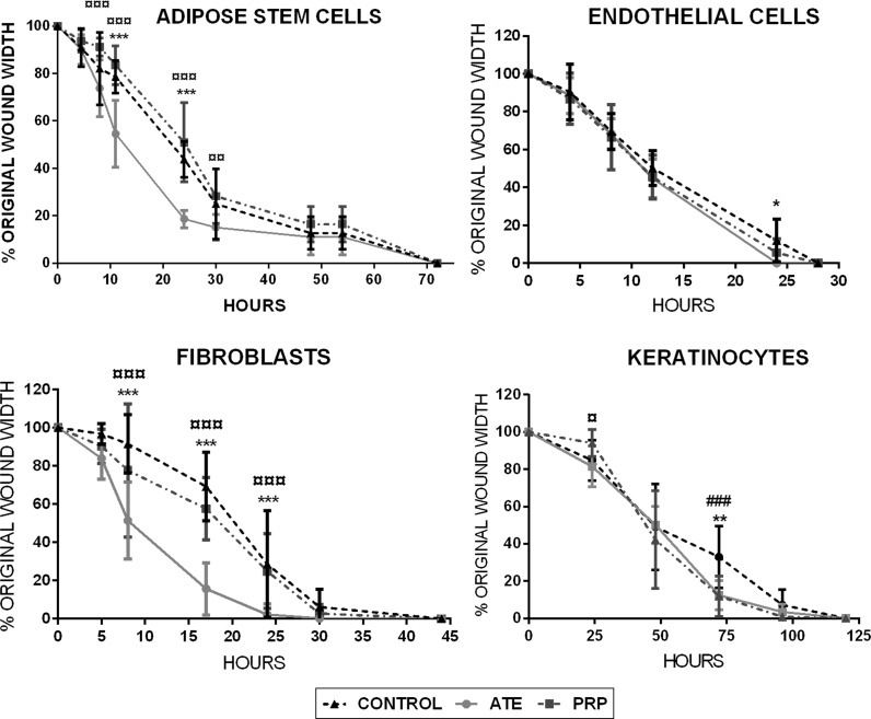
Fig. 3.
In vitro scratch wound assays presented in percentages of original wound width. Cell migration of the four studied cell types was measured under microscopy at determined time points until complete wound closure was achieved. In all treatment groups, adipose stem cells reached wound closure within 74 h, endothelial cells within 25 h, fibroblasts within 30 h and keratinocytes within 125 h. Results are depicted as mean ± SD. The statistical analysis was performed with two-way ANOVA with Tukey’s post-test. Differences were considered significant when p < 0.05*, p < 0.01** and p < 0.001***. Significances between ATE and control group are shown with (*), significances between PRP and control group with (#) and significances between ATE and PRP with (¤)

