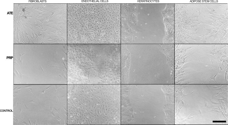
Fig. 4.
Phase contrast microscopic images of in vitro cell migration after scratch wound assays of fibroblasts, endothelial cells, keratinocytes and adipose stem cells at the 24 h timepoint. Cells were plated with the following counts: keratinocytes 300,000 cells/cm2, fibroblasts 100,000 cells/cm2, adipose stem cells 100,000 cells/cm2 and endothelial cells 150,000 cells/cm2. These cultures were treated with ATE, PRP or with their optimal cell culture medium, as control. Scale bar 100 µm

