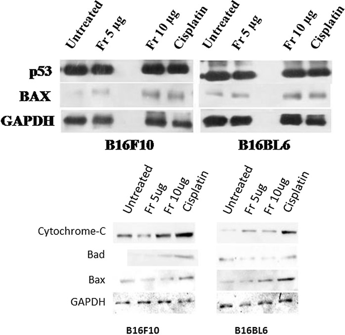
Fig. 7.
Protein expression (p53, Bax in the upper panel and Bad, cytochrome C in the lower panel) by B16F10 and B16BL6 cell in untreated condition, and after treatment with friedelane at 5, 10 μg and cisplatin (AT 10 µg/ml). Western blot images showed that proapoptotic protein levels increased after friedelane and cisplatin treatment

