Fig. 1.
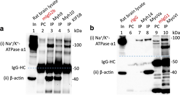
Interaction of multiple myosins with Na+/K+-ATPase α1 subunits expressed in rat brain. WT adult rat brain lysates (In, lane 1 in a and b) were precleared (PC) with indicated immunoglobulin isotypes (PC; lane2 = mIgG2b, lane 6 = rIgG and lane 9 = mIgG1) prior to immunoprecipitation (IP) using indicated antibodies (IP; lane 3 = myh9, lane 4 = myh10, lane 5 = KIF5B, lane 7 = Myh14, lane 8 = myoVa and lane 10 = myoVI). Loading of PC complexes in the gel preceded those of the IP complexes. Na+/K+-ATPase α1 subunits (i) were co-immunoprecipitated with myh9, myh10, KIF5B, myh14, myoVa and myoVI expressed in rat brain tissues. Co-immunoprecipitation of Na+/K+-ATPase α1 subunits by KIF5B served as a positive control. All the myosins assayed co-immunoprecipitated β-actin (ii). Denatured mouse IgG-HC (i.e., lanes 2–5, 9 and 10; panel (ii)), but not those of rabbit IgG (i.e., lanes 6–8) separated from their intact immunoglobulins (that is used for PC or IP) could be seen as this section was probed with mouse anti-β-actin antibodies

