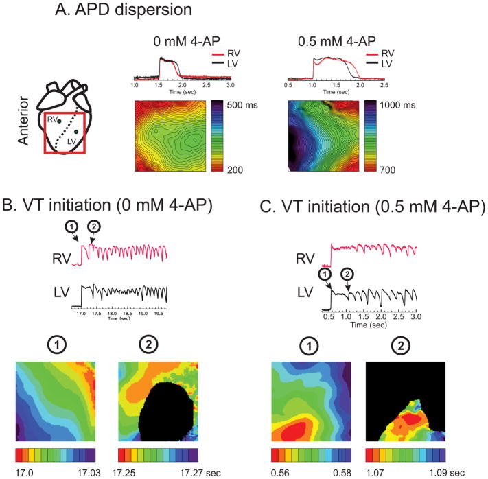
Figure 1.
A) APD maps under 4-AP. APD is longer in LV under normal conditions in LQT1 but 4-AP prolonged APDs (135% increase from 312 ± 63 to 729 ± 143 ms at 0.5 Hz) and flipped the APD gradient, resulting in longer APD in RV (n=4/5 hearts, Fisher exact test p < 0.05). B) RV initiation of pVT in LQT1 hearts (RV initiation 83% of pVT events, n=5/5 hearts, p < 0.0510). C) LV initiation of pVT in LV after 0.5 mM 4-AP (n=4/5 hearts, Fisher exact test p < 0.05). The black area indicates failed activation due to conduction block (Movies of activation are provided as supplemental material movie S1 and S2).

